- For Appointment: +91 89585 04460
- Mon-Sat (10am - 7pm)
- bhavnagulati8@gmail.com


Surgical Services
Laparoscopy
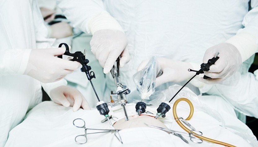
A laparoscopic is a process which involves placing small incisions in the velly or pelvis and utilising a camera for guidance. With a few microscopic abdominal surgeries, the laparoscope permits diagnostic or therapeutic procedures.
Modern surgical methods include laparoscopic surgery, often known as minimally invasive surgery, bandage surgery, or keyhole surgery. Comparing laparoscopic surgery to an exploratory laparotomy has a lot of benefits for the patient. Smaller incisions result in less pain, less bleeding, and quicker healing times. A laparoscope, a long fibre optic cable system that enables observation of the damaged area by snaking the cable from a more remote but more accessible position, is the crucial component.
Hysteroscopy
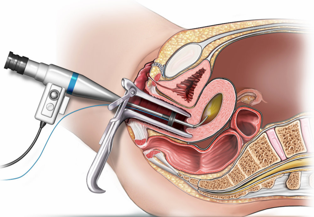
Hysteroscopy is a procedure that allows your doctor to look inside your uterus in order to diagnose and treat causes of abnormal bleeding. Hysteroscopy is done using a hysteroscope, a thin, lighted tube that is inserted into the vagina to examine the cervix and inside of the uterus. Hysteroscopy can be either diagnostic or operative.
Diagnostic hysteroscopy is used to diagnose problems of the uterus. Diagnostic hysteroscopy is also used to confirm results of other tests, such as hysterosalpingography (HSG). HSG is an X-ray dye test used to check the uterus and fallopian tubes.
Additionally, hysteroscopy can be used with other procedures, such as laparoscopy, or before procedures such as dilation and curettage (D&C). In laparoscopy, your doctor will insert an endoscope (a slender tube fitted with a fiber optic camera) into your abdomen to view the outside of your uterus, ovaries and fallopian tubes. The endoscope is inserted through an incision made through or below your navel.
Operative hysteroscopy is used to correct an abnormal condition that has been detected during a diagnostic hysteroscopy. If an abnormal condition was detected during the diagnostic hysteroscopy, an operative hysteroscopy can be performed at the same time, avoiding the need for a second surgery. During operative hysteroscopy, small instruments used to correct the condition are inserted through the hysteroscope.
Hysterectomy - Vaginal & Abdominal
Hysterectomy is the surgical removal of the uterus. It may also involve removal of the cervix, ovaries, fallopian tubes and other surrounding structures. Hysterectomy may be total (removing the body, fundus, and cervix of the uterus; often called "complete") or partial (removal of the uterine body while leaving the cervix intact; also called "supracervical").
Removal of the uterus renders the patient unable to bear children (as does removal of ovaries and fallopian tubes) and has surgical risks as well as long-term effects, so the surgery is normally recommended only when other treatment options are not available or have failed.
After a hysterectomy, you no longer have menstrual periods and cannot become pregnant. Hysterectomy is needed when the following symptooms occur:
- Uterine fibroids - Uterine fibroids are noncancerous growths in the wall of the uterus. In some women they cause pain or heavy bleeding.
- Heavy or unusual vaginal bleeding - Changes in hormone levels, infection, cancer, or fibroids can cause heavy, prolonged bleeding.
- Uterine prolapse - This is when the uterus slips from its usual place down into the vagina. This is more common in women who had several vaginal births, but it can also happen after menopause or because of obesity. Prolapse can lead to urinary and bowel problems and pelvic pressure.
- Endometriosis - Endometriosis happens when the tissue that normally lines the uterus grows outside of the uterus on the ovaries where it doesn't belong. This can cause severe pain and bleeding between periods.
- Adenomyosis - In this condition the tissue that lines the uterus grows inside the walls of the uterus where it doesn't belong. The uterine walls thicken and cause severe pain and heavy bleeding.
- Cancer (or precancer) of the uterus, ovary, cervix, or endometrium (the lining of the uterus). - Hysterectomy may be the best option if you have cancer in one of these areas.
Ovariotomy
An Ovariotomy (oophorectomy) is a surgical procedure to remove one or both of a woman's ovaries. The surgery is usually performed to prevent or treat certain conditions, such as ovarian cancer or endometriosis.
The surgery may just remove the ovaries, or it may be a part of a hysterectomy, which is the removal of the uterus and possibly some surrounding structures.
There are different reasons for an oophorectomy, including:
- treating abnormal tissue growth from endometriosis
- removing ovarian cysts, abscesses, or cancerous cells in the ovaries
- removing the source of estrogen, which may stimulate some cancer, such as breast cancer
Types of oophorectomy
Oophorectomy is a broad term for a medical procedure that removes one or both ovaries, but there are different types of oophorectomies that can be done.
- Unilateral oophorectomy - Removal of one ovary, usually done when a woman still wants to become pregnant.
- Bilateral oophorectomy - Removal of both ovaries, done to prevent disorders or spread of cancer cells.
- Salpingo oophorectomy - Removal of the fallopian tube along with the ovary, often to treat cancers or other disorders.
- Prophylactic oophorectomy - Also called a preventative oophorectomy, this procedure is done to reduce the risk of future diseases.
Dilation and Curettage (D and C) Evacuation
A dilation and curettage procedure, also called a D&C, is a surgical procedure in which the cervix (lower, narrow part of the uterus) is dilated (expanded) so that the uterine lining (endometrium) can be scraped with a curette (spoon-shaped instrument) to remove abnormal tissues. A suction D&C uses suction to remove uterine contents. This is sometimes called a dilation and evacuation (D&E).
Reasons for the procedure
- A D&C may be used as a diagnostic or therapeutic procedure for abnormal bleeding. A D&C may be performed to determine the cause of abnormal or excessive uterine bleeding, to detect cancer, or as part of infertility (inability to become pregnant) investigation.
- Causes of abnormal bleeding include the presence of abnormal tissues, such as fibroid tumors (benign tumors that develop in the uterus, also called myomas) polyps, or cancer of the endometrium or uterus. Tissues obtained from the D&C can be examined under a microscope. Abnormal uterine bleeding may also be due a hormone imbalance or disorder (particularly estrogen and progesterone) especially in women approaching menopause or after menopause.
- A D&C may be used following a miscarriage to remove the fetus and other tissues if they have not all been naturally passed. Infection or heavy bleeding can occur if these tissues are not completely removed. This type of D&C may also be called a surgical evacuation of the uterus or a D&E.
- Occasionally following childbirth, small pieces of the placenta (afterbirth) remain adhered to the endometrium and are not passed. This can cause bleeding or infection. A D&C may be used to remove these fragments so that the endometrium can heal properly.
Cervical Biopsy
A cervical biopsy is a surgical procedure in which a small amount of tissue is removed from the cervix. The cervix is the lower, narrow end of the uterus located at the end of the vagina. A cervical biopsy is a procedure to remove tissue from the cervix to test for abnormal or precancerous conditions, or cervical cancer. The cervix is the lower, narrow part of the uterus. It forms a canal that opens into the vagina.
Types of cervical biopsies
- Punch biopsy - In this method, small pieces of tissue are taken from the cervix with an instrument called “biopsy forceps.” Your cervix might be stained with a dye to make it easier for your doctor to see any abnormalities.
- Cone biopsy - This surgery uses a scalpel or laser to remove large, cone-shaped pieces of tissue from the cervix. You’ll be given a general anesthetic that will put you to sleep.
- Endocervical curettage (ECC) - During this procedure, cells are removed from the endocervical canal (the area between the uterus and vagina). This is done with a hand-held instrument called a “curette.” It has a tip shaped like a small scoop or hook.
Tubal Surgery (Tubal Ligation)
Tubal ligation is a surgical procedure that permanently closes or blocks your fallopian tubes. Every month, an egg leaves one of your ovaries (called ovulation). The egg moves through one of your fallopian tubes for a few days, waiting for sperm to come fertilize it. Pregnancy happens if a sperm cell meets up with one of your eggs, and the fertilized egg implants in your uterus.
When the fallopian tubes are blocked after a tubal ligation, sperm can't get to an egg and cause pregnancy. Tubal ligation is sometimes known as sterilization, female sterilization or “getting your tubes tied.”
Tubal ligation is surgery to block a woman's Fallopian tubes. A tubal ligation is a permanent form of birth control. After this procedure has been performed, an egg cannot move from the ovary through the tubes (a woman has two Fallopian tubes), and eventually to the uterus. Also, sperm cannot reach the egg in the Fallopian tube after ovulation (release of an egg from the ovary). Thus, pregnancy is prevented.
Myomectomy
A myomectomy is an operation to remove fibroids while preserving the uterus. For women who have fibroid symptoms and want to have children in the future, myomectomy is the best treatment option.
Myomectomy is very effective, but fibroids can re-grow. The younger you are and the more fibroids you have at the time of myomectomy, the more likely you are to develop fibroids again in the future. Women nearing menopause are the least likely to have recurring problems from fibroids after a myomectomy.
A myomectomy can be performed several different ways. Depending on the size, number and location of your fibroids, you may be eligible for an abdominal myomectomy, a laparoscopic myomectomy or a hysteroscopic myomectomy.
- Abdominal myomectomy lets your surgeon removes your fibroids through an open surgical cut in your lower belly.
- Laparoscopic myomectomy allows your surgeon toremove your fibroids through several small incisions. This may be done robotically. It’s less invasive and recovery is faster than with abdominal myomectomy.
- Hysteroscopic myomectomy requires your surgeon to use a special scope to remove your fibroids through your vagina and cervix.
Vulval cyst removal
Your gynecologist will use local anesthesia to eliminate pain. The surgical drainage involves making a small incision into the cyst and draining its contents. After draining the cyst, the doctor could place a small rubber tube in the incision to ensure that the cyst drains fully.
Repair of Perineal Tears
After vaginal delivery, the vagina, perineum, and anorectum are examined to identify and repair significant injuries. In particular, occult injury to the anal sphincter complex may occur at the time of an otherwise uncomplicated delivery and, if neglected, can contribute to anal and fecal incontinence. Even when recognized and repaired, persistent sphincter dysfunction is considered to be the most common cause of postpartum anal incontinence.
There are four degrees of tears that can occur during delivery:
- First degree tears involve the vaginal mucosa and connective tissue.
- Second degree tears involve the vaginal mucosa, connective tissue and underlying muscles.
- Third degree tears involve complete transection of the anal sphincter.
- Fourth degree tears involve the rectal mucosa.
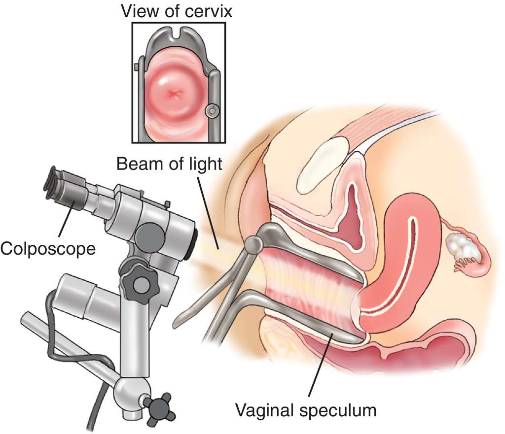
Colposcopy
A colposcopy is a type of cervical cancer test. It lets your doctor or nurse get a close-up look at your cervix - the opening to your uterus. It’s used to find abnormal cells in your cervix. Colposcopy is a procedure to closely examine your cervix, vagina and vulva for signs of disease. During colposcopy, your doctor uses a special instrument called a colposcope.
The procedure is usually performed if the results of a Pap smear (the screening test used to identify abnormal cervical cells) are unusual. A colposcope is a large, electric microscope with a bright light that enables your doctor to see your cervix more clearly and under magnification. If your doctor spots any abnormal areas, they will take a tissue sample (biopsy). The procedure to retrieve a tissue sample from inside the opening of the cervix is called endocervical curettage (ECC).
Why is colposcopy done?
Colposcopy is done when results of cervical cancer screening tests show abnormal changes in the cells of the cervix. Colposcopy provides more information about the abnormal cells. Colposcopy also may be used to further assess other problems:
- Genital warts on the cervix
- Cervicitis (an inflamed cervix)
- Benign (not cancer) growths, such as polyps
- Pain
- Bleeding
Sometimes colposcopy may need to be done more than once. It also can be used to check the result of a treatment.